
Tsinghua Scientists Grow Beating Heart from Stem Cells in Major Regeneration Breakthrough
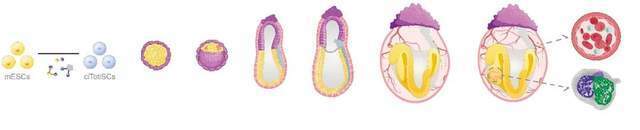
The team’s workflow involves a two-stage process: first using chemical methods to intervene in mouse stem cells, and then placing the modified cells into a culture environment for gradual expansion. These cell populations spontaneously aggregate into a whole, simulating the differentiation trajectory of a normal embryo. They progressively develop from pluripotent stem cells into various cell types, eventually giving rise to the primitive shapes of organ tissues.
After a 12-day culture cycle, some of the models even developed rhythmically beating heart tissue. Ding noted that while the developmental process of normal mouse embryos is well understood by the scientific学界 (academic community), whether *in vitro*-induced cell lines can autonomously complete all developmental stages from start to finish—and how far they can advance—was entirely unpredictable before the experiment.
Should this research eventually overcome current bottlenecks, Ding envisions profound applications: potentially reviving extinct species or even achieving human “biological restoration.” Yet the current limitations are equally apparent—the embryo models created in this study survived for a maximum of just 12 days. A more pressing question remains: how vast is the chasm between a cluster of beating heart cells and a fully functional, active mouse?

Chemically induced pluripotent stem cells evolve into mouse embryo morphology *in vitro*, initiating the early stages of organ development. Image source: Tsinghua University official website
Embryonic Systems Capable of Pulsation
The primary research subjects for this experiment were mouse embryonic stem cell lines. Researchers initially treated these cells with chemical agents and transferred them into culture plates. Capable of automatically aggregating without artificial intervention, the cells self-assemble into spherical structures, evolving sequentially much like real embryos. These resulting cell groups are known as embryo model bodies.
By the fourth day, most of the cell clusters closely resembled mouse embryos at three to four days post-conception. When the tenth day arrived, preliminary forms of neural and intestinal tissues began to appear in a small number of embryo bodies, alongside the formation of vascular networks and the onset of hematopoietic (blood-producing) functions. Entering the twelfth day, researchers discovered heart organs differentiated into left and right atria and ventricles within individual embryo bodies, complete with blood cells inside the cardiac chambers. Evaluated solely by the heart, the developmental level was equivalent to that of a 10.5-day normal mouse embryo.
The critical hinge of this research lies in the selection and processing of the initial stem cells. In normal mouse development, embryos sequentially form two auxiliary structures: the placenta and the yolk sac, whose functions are to supply nutrients and developmental instruction signals to the embryo body. The yolk sac is also the initial site where blood formation is achieved. The fetal trunk, placenta, and yolk sac all originate from the incremental division of a single fertilized egg. Although untreated stem cells possess multi-directional differentiation potential and can grow into various internal cell types, they lack the capacity to generate the placenta and yolk sac. What Ding pursued was precisely this kind of primordial stem cell—one equivalent to the totipotent foundational cell of a fertilized egg.
However, up to now, whether through natural reproduction or nuclear transfer cloning, generating new life has inevitably required germ cells such as eggs or sperm. Ding’s approach reversed this logic: using chemical substances to wind back the developmental timeline of stem cells to the “zero point” of life, and then allowing this group of cells to autonomously sprout and evolve according to their internal instructions.
To achieve this goal, Ding’s team spent years. Beginning in 2015, Ding led a search for the “magic reagents” capable of accomplishing this feat. In 2017, the team successfully found several chemical molecules that could guide some stem cells to exhibit properties closer to totipotent stem cells, possessing key characteristics similar to a fertilized egg at the source of life. However, this discovery was not published at the time because Ding observed that this special cellular state was fleeting and difficult to maintain stably through artificial means.
It was not until 2022 that Ding’s team revealed the true identity of these “magic reagents” in *Nature*. Their molecular composition includes three highly active small chemical molecules that can precisely control intracellular gene expression, safely resetting stem cells to a naive state. Researchers fluorescently labeled a small number of these cells and injected them into the early embryos of normal mice. As the embryos grew into mice, fluorescently labeled cells were detected within the tissues of the fetal trunk, placenta, and yolk sac, fully demonstrating that these cells played a role in the growth of all three vital structures.
However, those cells were previously implanted into real mouse embryos, completing the entire developmental journey alongside a host embryo. The new experiments differ: by integrating an additional molecular substance onto the original “reagents,” cells modified in this way could independently evolve into the three-dimensional structure of an embryo body using only themselves.
Ding’s team was not the first to cultivate pulsating mouse embryo bodies using stem cells. In 2022, two research teams from the Weizmann Institute of Science in Israel and the University of Cambridge in the UK achieved this feat using an “assembly” approach. These researchers prepared three different types of stem cell cultures—one dedicated to evolving into the mouse fetal body, one specialized in forming placental tissue, and one specialized in generating yolk sac structures—and then mixed and fused the three cell types to build an embryo body.
Ding pointed out that embryo bodies constructed via this “assembly” method bypass the blastocyst stage, making it impossible for them to implant into a uterine environment. For an embryo to develop further, it must undergo implantation—embedding into the endometrium—which naturally occurs during the blastocyst stage. The embryo bodies cultivated by Ding’s team, by contrast, completely experience the blastocyst stage, theoretically possessing the feasibility of being returned to a maternal uterus.
Ding noted that among the embryo body models cultivated by various research groups currently, heart development remains at an early stage, and no essential divergence between the two experimental approaches can yet be observed. However, he believes his team’s technical route can ultimately go deeper. In his theoretical assessment, the process of embryonic development amplifies minor defects step-by-step. The “assembly” method requires not only that all three cell types meet specification requirements, but also flawless assembly craftsmanship; if any single link goes wrong, the impact of that problem will grow exponentially as development continues. Nonetheless, he candidly admitted that the “assembly” approach might be easier for achieving short-term research breakthroughs currently.

In 2026, Ding Sheng (front row, center) poses for a group photo with members of his scientific research team. Image source: Tsinghua University official website
Why Further Growth Stalls
The longer the culture time is extended, the more sharply the number of embryo bodies capable of maintaining normal development drops. By the critical milestone of the fourth day, researchers identified cell groups with better developmental progress and transferred them to the next culture phase. Among these embryo bodies, about one-fifth maintained normal developmental progress on the seventh day; by the eighth day, this proportion dropped below 10 percent; and by the tenth day, embryo bodies with clearly distinguishable head and tail structures became scarce, falling below 1 percent. By the twelfth day, only three embryo body samples completed this developmental stage.
Research data exposed several key anomalies. During observations on the fourth day, the number of cells constructing the yolk sac within some cell clusters was excessive, while the number forming the placenta was insufficient. Ding also pointed out that within the embryo bodies existed another batch of cells whose developmental pace was uncoordinated with the rhythm of the overall system. In his analytical framework, these deviations could all evolve into potential factors hindering the embryo bodies from progressing further.
He compared the evolutionary process of an embryo to a large-scale group dance performance. Cells must undergo differentiation and transition strictly in chronological order, generating corresponding tissue structures while continuously releasing biological signals to surrounding cells to achieve precise mutual coordination. “The timing must be precise; it cannot be early, it cannot be delayed, and it certainly cannot fall out of step,” he explained. Cells with misaligned developmental timestamps—significantly faster or slower than expected—acted like dancers missing a beat, causing the rhythm of surrounding cells to descend into chaos and leading to errors multiplying and accumulating exponentially.
Why do embryo bodies become increasingly difficult to maintain normal developmental progress in later stages? Ding proposed two possible directions for explanation. The first hypothesis involves the initial stem cells themselves. Although these stem cells, dialed back to a “primitive state” via chemical means, possess most of the characteristics of early totipotent stem cells, they still retain certain lingering traits from their starting point, which may create obstacles on the path of the embryo body developing into a complete individual.
The second hypothesis points to the limitations of external culture conditions. Inside the uterine environment, an embryo relies on the mother’s blood supply to obtain oxygen and nutrients, whereas the laboratory lacks this maternal biological system and must rely on artificial compensation. Ding’s judgment is that such simulated alternatives are not yet comprehensive or complete enough, likely serving as a major factor making it difficult for embryo bodies to sustain stable development.
It remains inconclusive whether the impact of one factor dominates or if both collaborate to produce the result. The team has also attempted to implant mouse embryo bodies into a mouse uterine environment, though the conclusions of those experiments have yet to be publicly released. Ding stated that the embryo bodies can indeed complete implantation within the uterus, causing the uterine wall to thicken and swell, but within ten days of development, the embryo bodies are absorbed and dissolved by the uterine tissue. Ding believes this reflects the operation of natural selection mechanisms inherent within biological systems, whereby embryos whose developmental status fails to reach ideal standards are naturally eliminated.
Regarding the topic of reviving extinct species, Ding expressed that “this work does not necessarily have to be undertaken by our team.” His own primary interests lean more toward utilizing this technological means to conduct deep academic research at the macro level of biological evolution, or even imagining what shape humanity will evolve into tens or hundreds of thousands of years from now. As for the concept of “human resurrection,” he believes that from the perspective of scientific realization, it is essentially identical to nuclear transfer cloning, involving profound moral and ethical issues within the current ethical and social context.
If such cutting-edge technologies are applied to humans, the setting of ethical boundaries must be clear and explicit. The International Society for Stem Energy Research (ISSCR) explicitly stipulates in its latest ethical guidelines issued in 2025 that implanting embryo body models containing human stem cells into the uterus of any human or animal is strictly prohibited. Within China, the 2025 *Ethical Guidelines for Human Organoid Research and Development* similarly enforce this ban clearly.
Ding’s team wrote in the concluding section of their research paper that alongside the continuous optimization and upgrading of such mouse embryo bodies, related scientific research methodologies may be explored in human cell experiments in the future. However, this will inevitably demand the establishment of strict regulatory ethical framework mechanisms to prevent their abuse in the field of reproductive propagation.
As for when the first live mouse born through this technology might make its debut, Ding cannot yet provide a clear timeline expectation. “This remains an open scientific puzzle, rather than a purely engineering technical problem,” he said, noting that the mechanisms of numerous issues remain unclear, and scientific research can only move forward through continuous experimentation and defect correction.